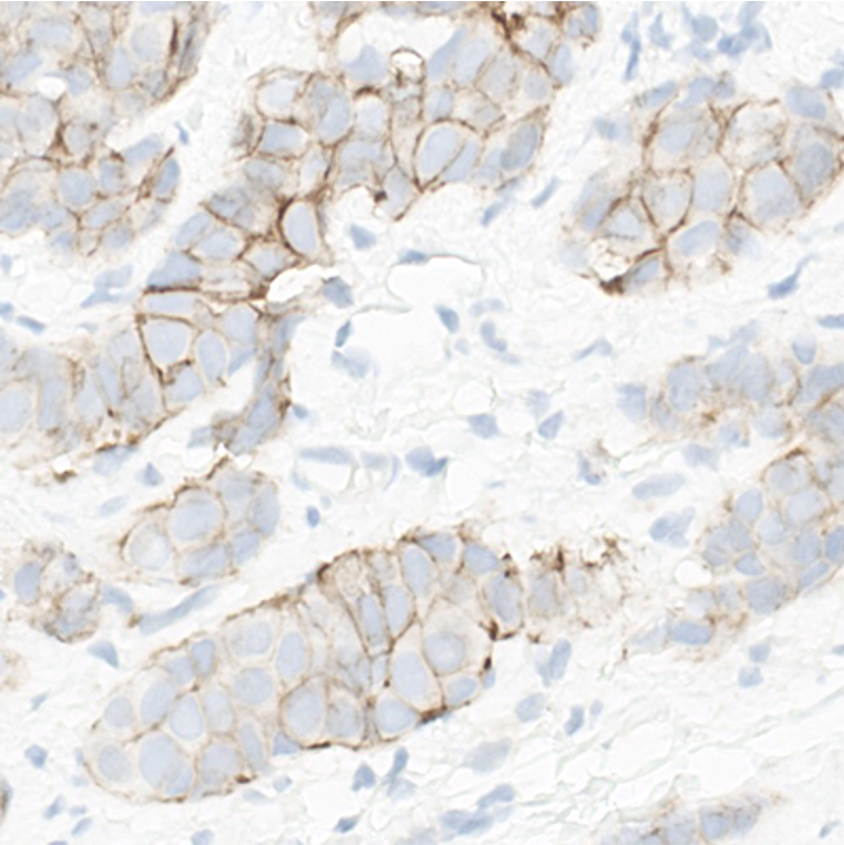
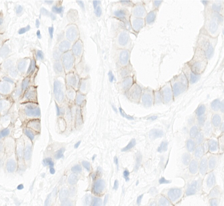








Your selected HER2 IHC score is aligned with the consensus score provided by the Pathology Faculty.
Consensus score: 0
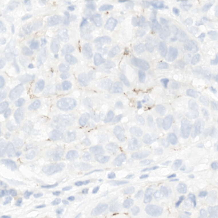
This is a challenging case where about 10% of tumor cells display a faint/barely perceptible incomplete membranous staining. In such borderline cases, screening the slide multiple times from low (4X) to high magnification (20X-40X) is needed.
As this tumor has a poorly differentiated morphology, repeating the IHC on a different specimen, such as an excision, might be considered. A larger tissue sample from high-grade tumors might show a different IHC result from the one obtained from a tissue biopsy.
Your selected HER2 IHC score is not aligned with the consensus review provided by the Pathology Faculty.
Consensus score: 0
This is a challenging case where about 10% of tumor cells display a faint/barely perceptible incomplete membranous staining. In such borderline cases, screening the slide multiple times from low (4X) to high magnification (20X-40X) is needed.
As this tumor has a poorly differentiated morphology, repeating the IHC on a different specimen, such as an excision, might be considered. A larger tissue sample from high-grade tumors might show a different IHC result from the one obtained from a tissue biopsy.
Your selected HER2 IHC score is aligned with the consensus score provided by the Pathology Faculty.
Consensus score: 0
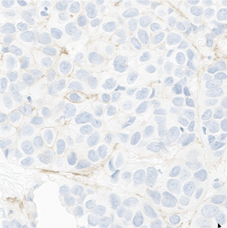
Less than 10% of tumor cells display a faint/barely perceptible and mainly incomplete membranous staining. This case could be challenging since the specifically stained tumor cells are scattered while multiple tumor cells only present a non-specific basal staining.
Your selected HER2 IHC score is not aligned with the consensus review provided by the Pathology Faculty.
Consensus score: 0
Less than 10% of tumor cells display a faint/barely perceptible and mainly incomplete membranous staining. This case could be challenging since the specifically stained tumor cells are scattered while multiple tumor cells only present a non-specific basal staining.
Your selected HER2 IHC score is aligned with the consensus score provided by the Pathology Faculty.
Consensus score: 0
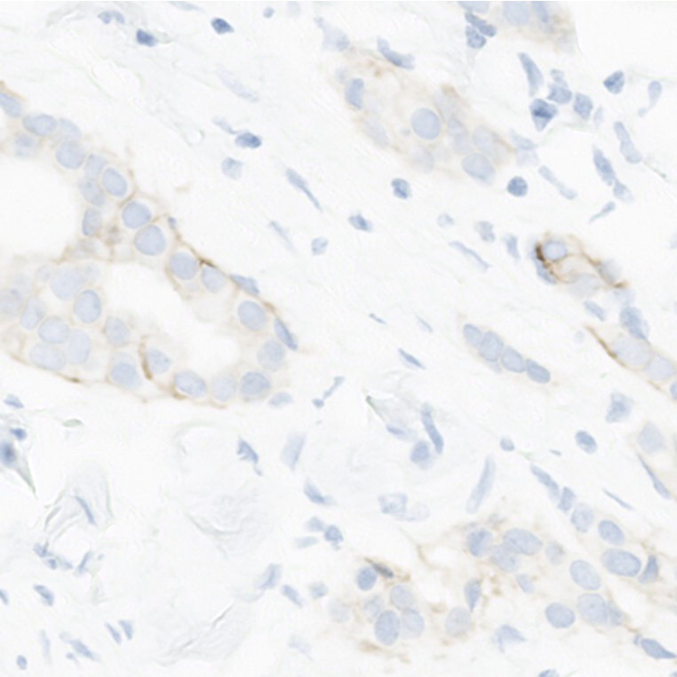
This is an interesting case where several tumor cells exhibit a faint and rarely moderate incomplete membranous staining. However, when carefully examined, the percentage of these stained tumor cells is less than 10%.
The strong staining intensity might create a false impression of a higher number of stained tumor cells.
Your selected HER2 IHC score is not aligned with the consensus review provided by the Pathology Faculty.
Consensus score: 0
This is an interesting case where several tumor cells exhibit a faint and rarely moderate incomplete membranous staining. However, when carefully examined, the percentage of these stained tumor cells is less than 10%.
The strong staining intensity might create a false impression of a higher number of stained tumor cells.
Your selected HER2 IHC score is aligned with the consensus score provided by the Pathology Faculty.
Consensus score: 2+
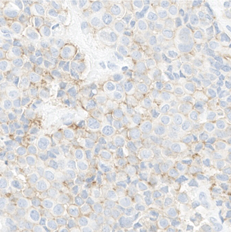
This is an interesting case where more than 10% of tumor cells display a moderate to strong incomplete basolateral or lateral membranous staining. The faint staining might mislead some to score this case 1+.
Your selected HER2 IHC score is not aligned with the consensus review provided by the Pathology Faculty.
Consensus score: 2+
This is an interesting case where more than 10% of tumor cells display a moderate to strong incomplete basolateral or lateral membranous staining. The faint staining might mislead some to score this case 1+.
Your selected HER2 IHC score is aligned with the consensus score provided by the Pathology Faculty.
Consensus score: 1+
This an interesting case with several tumor cells displaying a faint/barely perceptible and incomplete membranous staining while some are expressing a strong basolateral or lateral staining. The high intensity staining in less than 10% of tumor cells keeps this case at a lower score.
As this tumor has a poorly differentiated morphology and micropapillary features, repeating the IHC on a different specimen, such as an excision, might be considered. A larger tissue sample from high-grade tumors might show a different IHC result from the one obtained from a tissue biopsy.
Your selected HER2 IHC score is not aligned with the consensus review provided by the Pathology Faculty.
Consensus score: 1+
This an interesting case with several tumor cells displaying a faint/barely perceptible and incomplete membranous staining while some are expressing a strong basolateral or lateral staining. The high intensity staining in less than 10% of tumor cells keeps this case at a lower score.
As this tumor has a poorly differentiated morphology and micropapillary features, repeating the IHC on a different specimen, such as an excision, might be considered. A larger tissue sample from high-grade tumors might show a different IHC result from the one obtained from a tissue biopsy.